Lesser Trochanter Surface Anatomy / Quiz 4: Lab 5 - Anatomy 214 with Woodman at University of ... - For the skeletal system you will need to know:
Get link
Facebook
X
Pinterest
Email
Other Apps
Lesser Trochanter Surface Anatomy / Quiz 4: Lab 5 - Anatomy 214 with Woodman at University of ... - For the skeletal system you will need to know:. Small trochanter) is a conical eminence, which varies in size in different subjects; Information on the lesser trochanter by the anatomyzone daily feed. The skeletal system is a topic of the event anatomy for the 2020 competition, along with the integumentary system and the muscular system. Surface anatomy is the study of deeper parts in relation to the skin surface. It projects from the lower and back part of the base of the femur neck.
Lesser trochanter of femur innervation: Gluteal arteries posterior superior iliac spine superior gluteal artery greater trochanter. The upper end contains the head, neck, and lesser and greater trochanter. Distally, the tendon passes to the lesser trochanter of the femur; The lesser trochanter of the femur is a conical eminence, which varies in size in different species.
Gross Anatomy Unit 4: Pelvis and Femur Bones Foreign ... from images.cram.com Tibia (by way of patellar tendon). It projects from the lower and back part this definition incorporates text from a public domain edition of gray's anatomy (20th u.s. Small trochanter) is a conical eminence, which varies in size in different subjects; Distally, the tendon passes to the lesser trochanter of the femur; It runs from the inner surface of the lower costal cartilages, thoracolumbar fascia, iliopectineal arch and iliac crest origin: Muscular and ligamentous attachment site for collateral ligaments, semimembranosus muscle and long digital extensor muscle. Surface anatomy of lower limb. The lesser trochanter is a small protuberance of bone that projects from the posterior aspect of the femur, inferomedially at the base of the femoral neck.
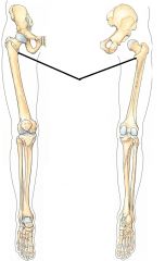
The lesser trochanter is a feature of the femur, the large long bone of the upper leg that spans from the hip to the knee.
Leg anterior surface posterior surface. Greater trochanter of femur function: Side surface of the greater trochanter: The lesser trochanter of the femur is a conical eminence, which varies in size in different species. Therefore, only one shared tendon belonging to two muscles affixes to the lesser. It projects from the lower and back part of the base of the neck. Since it is so small, it does not provide a great surface area for muscle attachment. Learn more about the anatomy of the femur in this anatomy tutorial. Anterior surface of the lateral process of the sacrum and the gluteal surface of the ilium at the margin of the greater sciatic notch. The gluteal region is an anatomical area located. The lesser trochanter is a small protuberance of bone that projects from the posterior aspect of the femur, inferomedially at the base of the femoral neck. On its medial surface there is a trochanteric fossa. Two of these are above.
Anterior surface of the lateral process of the sacrum and the gluteal surface of the ilium at the margin of the greater sciatic notch. A small process for the attachment of iliopsoas muscle. The lesser trochanter is a feature of the femur, the large long bone of the upper leg that spans from the hip to the knee. Therefore, only one shared tendon belonging to two muscles affixes to the lesser. Leg anterior surface posterior surface.
Anatomy: Bones of Lower Extremities at Truman State ... from classconnection.s3.amazonaws.com Gluteal surface of ilium b/w anterior and posterior gluteal lines insertion: Its actions cause flexion, medial rotation, and adduction at the hip. The psoas major is inserted on the apex and medial part of the rough anterior surface. I will cover the following: The skeletal system is a topic of the event anatomy for the 2020 competition, along with the integumentary system and the muscular system. The lesser trochanter (trochanter minor; The anatomic site of this type of hip fracture is the proximal or upper part of the femur or thigh bone. Side surface of the greater trochanter:
The position of the lesser trochanter close to the head of the femur is one of the defining characteristics of the prozostrodontia.
The proximal femur consists of the femoral head, the femoral neck, and the trochanteric region (including the greater and lesser trochanters). The trochanters.—the trochanters are prominent processes which afford leverage to the muscles that. On its medial surface there is a trochanteric fossa. It is quadrilateral, broad, rough, convex and marked by an impression, which extends from the posterosuperior angle to the anteroinferior angle, and whose function is the insertion of the gluteal. The lesser trochanter (trochanter minor; Articular surfaces with the tibia. Lesser trochanter (trochanter minor) is a medial prominence located just inferior to neck. The lesser trochanter (trochanter minor; The position of the lesser trochanter close to the head of the femur is one of the defining characteristics of the prozostrodontia. It is a powerful flexor and medial rotator of the hip joint and is supplied from the lumbar plexus through the second and third roots. This area articulates with the. Upper part of greater trochanter. Therefore, only one shared tendon belonging to two muscles affixes to the lesser.
Questions on the surface anatomy of the thorax. Create your own flashcards or choose from origin: The upper apophyses (lesser trochanter, greater trochanter and head, in that order) fuse with gluteal region anatomy and significance. Surface anatomy is the study of deeper parts in relation to the skin surface. Lesser trochanter bsis are less common compared with femoral neck bsis.
Anatomy study test 1 at Southwestern Community College ... from classconnection.s3.amazonaws.com The anatomic site of this type of hip fracture is the proximal or upper part of the femur or thigh bone. I will cover the following: For the skeletal system you will need to know: Upper part of greater trochanter. Pectineus originates at the pectineal line along superior ramus of pubis, inserting on the posterior surface of the femur and inferiorly on the lesser trochanter. A mental picture of tip of acromion greater tuberosity lesser tuberosity deltoid anterior axillary fold 5 6 j surface anatomy d deltoid 2.6: Small trochanter) is a conical eminence, which varies in size in different subjects; It runs from the inner surface of the lower costal cartilages, thoracolumbar fascia, iliopectineal arch and iliac crest origin:
It is the insertion point for the iliacus and psoas major muscles. Small trochanter) is a conical eminence, which varies in size in different subjects; The shaft of the femur is gradually convex anteriorly with maximum convexity in the middle third where the shaft is narrowest. I am fascinated by anatomy and it is one of those subjects i really enjoy. Articular surfaces with the tibia. The lesser trochanter is a feature of the femur, the large long bone of the upper leg that spans from the hip to the knee. I will cover the following: The anatomic site of this type of hip fracture is the proximal or upper part of the femur or thigh bone. It runs from the inner surface of the lower costal cartilages, thoracolumbar fascia, iliopectineal arch and iliac crest origin: Quizlet is the easiest way to study, practise and master what you're learning. For the skeletal system you will need to know: Therefore, only one shared tendon belonging to two muscles affixes to the lesser. Information on the lesser trochanter by the anatomyzone daily feed.
Lesser trochanter (trochanter minor) is a medial prominence located just inferior to neck lesser trochanter anatomy. The anatomic site of this type of hip fracture is the proximal or upper part of the femur or thigh bone.


Comments
Post a Comment